-
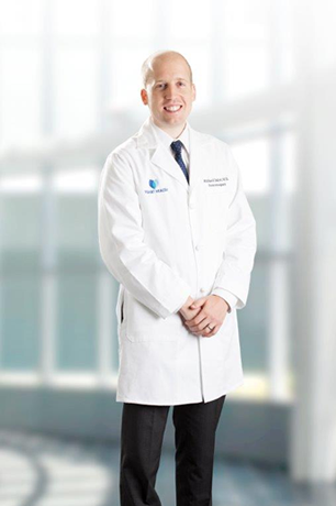 Welcome to the website ofDr. Richard DalyaiCerebrovascular & Endovascular NeurosurgeonDr. Dalyai is a vascular neurosurgeon and the Surgical Director of Stroke at Vidant Medical Center.
About Dr. Dalyai
Cerebrovascular and Endovascular Neurosurgeon
Experience:
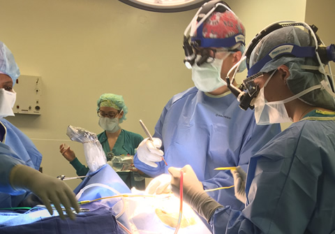
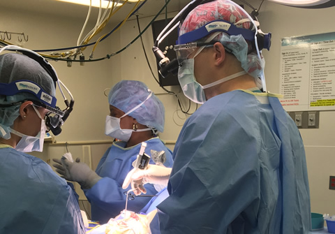
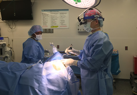
Dr. Richard T Dalyai is a vascular neurosurgeon and the Surgical Director of Stroke at Vidant Medical Center in Greenville, North Carolina. Dr. Dalyai is also adjunct faculty at the Brody School of Medicine at East Carolina University. He completed his neurosurgery residency at the Thomas Jefferson University Hospital working extensively under Dr. Robert Rosenwasser, one of the first neurosurgeons dual-trained in interventional neuroradiology procedures and an international leader in cerebrovascular neurosurgery. During his residency, Dr. Dalyai completed a clinical fellowship in endovascular and cerebrovascular neurosurgery, also working with Dr. Pascal Jabbour, a pioneer in aneurysm “pipeline” flow diversion, and Dr. Stavropoula Tjoumakaris, an expert in subarachnoid hemorrhage and cerebral arteriovenous malformations. Because of his extensive training, Dr. Dalyai has a unique dual-trained cerebrovascular skillset to treat infants, children, and adults with aneurysms, arteriovenous malformations, and subarachnoid hemorrhage using minimally invasive techniques to allow for the best possible outcomes.
Biography:
Richard T Dalyai, MD, joined the Department of Neurosurgery at Vidant Medical Center in 2016 as a fellowship dual-trained endovascular and cerebrovascular neurosurgeon. Dr. Dalyai is the Surgical Director of Stroke and also adjunct faculty with the Brody School of Medicine of East Carolina University in Greenville, NC. In Boston, he graduated from Tufts University School of Medicine in 2009. In Philadelphia, he completed his neurosurgery residency Thomas Jefferson University Hospital in 2016. He completed a fellowship in endovascular and cerebrovascular neurosurgery at the Thomas Jefferson University Hospital.
Dr. Dalyai treats all vascular neurosurgical disorders and specializes in the treatment of cerebral aneurysms, arteriovenous malformations, and dural arteriovenous fistulas. He is extensively published in subarachnoid hemorrhage, stereotactic radiosurgery, and ischemic stroke. He uses endovascular and open surgical techniques in the treatment of all vascular neurosurgical disorders in children and adults including carotid artery stenting and endarterectomy. He also treats spinal conditions cervical and lumbar disc herniations, spinal stenosis, and spondylisthesis.
Dr. Dalyai is the surgical director of stroke at Vidant Medical Center. As a fellow at Jefferson Hospital for Neuroscience, he worked with the Joint Commission to become a Comprehensive Stroke Center. Under the leadership of Dr. Robert Rosenwasser, Jefferson Neuroscience Institute pioneered the use a telehealth to optimize the system of ischemic stroke care.
Dr. Dalyai treats many conditions, including:
- Aneurysm
- Arteriovenous Malformations (AVMs)
- Carotid-Cavernous Sinus Fistula (CCF)
- Carotid Stenosis
- Dural AVFs
- Intracerebral Hemorrhage
- Ischemic Stroke
- Subarachnoid Hemorrhage
- Vasospasm
Make Appointment
Click here to download Dr. Dalyai's CV.
Dr. Dalyai is one of North Carolina's top fellowship trained Cerebrovascular Neurosurgeons and was trained at Thomas Jefferson University Hospital.
Educational Videos
Operative Videos
Frequently Asked Questions
A: A cerebral aneurysm (also referred to as intracranial or intracerebral aneurysm) is a weak or thin spot on a blood vessel in the brain that balloons out and fills with blood. Sometimes a bulging aneurysm can put pressure on a nerve or surrounding brain tissue; however, usually the major concern with cerebral aneurysms is that they may rupture. A ruptured aneurysm can pour blood or hemorrhage into the surrounding brain tissue. Some aneurysms, especially those that are tiny, do not cause any problems and are at a low risk of bleeding.
A: Brain aneurysms can develop in anyone, at any age. They are more common in adults than in children and slightly more common in women than in men. People with certain inherited disorders (such as polycystic kidney disease and connective tissue disorders) are also at a higher risk.
Almost all cerebral aneurysms have the potential to rupture and cause bleeding within the brain; however, some aneurysms are much more likely to rupture or cause other problems than others. The incidence of reported ruptured aneurysm is about 10 in every 100,000 persons per year (about 30,000 individuals per year in the U.S.), most commonly in people between ages 30 and 60 years. Possible risk factors for rupture include smoking tobacco, high blood pressure, alcohol abuse, and drug abuse (particularly cocaine).
A: Aneurysms may rupture and bleed around the surrounding brain tissue (called a subarachnoid hemorrhage), which can lead to hemorrhagic stroke, permanent nerve damage, or death. Once it has ruptured, the cerebral aneurysm has a very high chance of rupturing again. The high risk of re-rupture is why we quickly work to treat ruptured aneurysms. Another potential complication a ruptured aneurysm is hydrocephalus, in which the excessive buildup of cerebrospinal fluid in the skull dilates fluid pathways called ventricles that can swell and press on the brain tissue. Another delayed ccomplication of subarachnoid hemorrhage is called vasospasm, in which other blood vessels in the brain contract and limit blood flow to vital areas of the brain. This reduced blood flow can also cause stroke or tissue damage.
A: Most cerebral aneurysms do not show outward symptoms until they either become very large or burst. Small, unchanging aneurysms generally will not produce symptoms, whereas a larger aneurysm that is steadily growing may press on nerves, especially those relating to functions of the eye and vision.
When an aneurysm hemorrhages, a patient may experience a sudden and extremely severe headache, double vision, nausea, vomiting, stiff neck, and/or loss of consciousness. Individuals usually describe the headache as “the worst headache of my life” and it is generally different in severity and intensity from other headaches people may experience. “Sentinel” or warning headaches may result from an aneurysm that “leaks” for days to weeks prior to rupture. Only a minority of individuals have a sentinel headache prior to aneurysm rupture.
Other signs that a cerebral aneurysm has burst include nausea and vomiting associated with a severe headache, a drooping eyelid, sensitivity to light, and change in mental status or level of awareness. Some individuals may have seizures. Individuals may lose consciousness briefly or go into prolonged coma. People experiencing this “worst headache,” especially when it is combined with any other symptoms, should seek immediate medical attention.
A: Not all cerebral aneurysms burst, and some have a very low chance of ever causing problems. Some people with very small aneurysms may be monitored with MRI/A to detect any growth or onset of symptoms. Each patient and aneurysm is unique, and considerations for treating an unruptured aneurysm include the type, size, and location of the aneurysm; risk of rupture; the individual’s age, health, and personal and family medical history; and the risk of treatment.
Two surgical options are available for treating cerebral aneurysms. The first and most traditional is microvascular clipping which involves cutting off the flow of blood to the aneurysm. Under anesthesia, a section of the skull is removed and the aneurysm is located. The neurosurgeon uses a microscope to isolate the blood vessel that feeds the aneurysm and places a small, metal, clothespin-like clip on the aneurysm’s neck, halting its blood supply. The clip remains in the skull and prevents the risk of future bleeding. The piece of the skull is then replaced and the scalp is closed. Clipping has been shown to be highly effective, depending on the location, shape, and size of the aneurysm. In general, aneurysms that are completely clipped surgically do not return.
A second surgical option is endovascular coil embolization. Once under anesthesia, the neurosurgeon inserts a hollow plastic tube (a catheter) into an artery in the upper leg and threads it, using angiography, through the great vessels in the neck and to the site of the cerebral aneurysm. Once the catheter is in place, detachable coils (spirals of platinum wire) are released into the aneurysm. The coils fill the aneurysm and block it from the cerebral circulation, which effectively obliterates and destroys the aneurysm. This procedure may need to be performed more than once during the person’s lifetime.
Both of these surgical options carry some risk to the patient (such as possible damage to other blood vessels, the potential for aneurysm recurrence and rebleeding, and the risk of post-operative stroke).
A: There are several factors that weigh on the decision of the type of treatment recommended including your age and medical condition, as well as size, shape, and location of the aneurysm. With the new technology, many patients are able to be treated by endovascular aneurysm coiling that previously were unable to be treated; however, for many aneurysms clipping is the best modality.
A: Approximately 1½ – 3 hours.
A: Approximately 4 – 5 hours.
A: General endotracheal anesthesia is always used for aneurysm clipping procedures and most endovascular coiling procedures in which you are “asleep”. An attending anesthesiologist who is experienced in neuro-anesthesia is present at all times.
A: The duration of the hospital stay is very different for patients with ruptured versus unruptured aneurysms. Patients with unruptured aneurysms typically have a shorter hospital stay of approximately 2 – 3 days depending on the method of treatment. For patients who have suffered a ruptured aneurysm (or subarachnoid hemorrhage) length of stay is variable and is dependent on the condition of the patient on arrival and severity of the bleed. In patients with severe subarachnoid hemorrhage, hospitalization may be from 14 days to 1 month.
A: In patients where there is more than 1 family member diagnosed with an aneurysm, it is recommended that all family members should consult with their physicians to receive non-invasive screenings, such as MRA or CTA.
A: For most aneurysm clip surgery incisions heal in about 6 weeks. In some patients we are able to use dissolvable sutures which takes several weeks, in other’s staples are used and will be removed after two weeks in the clinic. After endovascular aneurysm coiling, there is quick healing of the puncture site in the groin.
A: Most aneurysms clip patients are off all pain medications within 1 – 3 weeks. Other medications, for instance, anti-hypertensive drugs, will be modulated by your primary care physician. After endovascular aneurysm coiling, selected patients may be on “blood-thinners”, such as Plavix or aspirin.
A: A major risk factor for developing aneurysms is smoking tobacco. There are medical conditions such as polycystic kidney disease, fibromuscular dysplasia, or any other type of elastic tissue disorder, where continued screening throughout your lifetime may be indicated. For most patients though, it is unlikely you will develop a new aneurysm.
A: Yes, all titanium clips and platinum coils we use are MRI compatible.
A: Patients with unruptured aneurysms usually should wait 2 weeks after the procedure before driving. Patients who have had unruptured aneurysms treated with endovascular coiling typically can drive sooner.
A: This is different for each patient, their medical condition before surgery, and, of course, the demands of their specific job.
A: This is not very well known. Aneurysms may slowly enlarge over time, but we cannot exactly predict the rate of growth. Some aneurysms, typically smaller aneurysms, do not change over a lifetime.
A: This depends on your medical history and aneurysm characteristics as well as treatment. After coiling, imaging follow-up (MRA, angiography) is necessary to confirm persistent occlusion upto 5 years. For aneurysm clipping, most patients do not need further follow up imaging.
A: This depends on your medical history and aneurysm characteristics as well as treatment. After coiling, imaging follow-up (MRA, angiography) is necessary to confirm persistent occlusion upto 5 years. For aneurysm clipping, most patients do not need further follow up imaging.
A: Cerebral arteriovenous malformations (or AVMs) are abnormal direct connection between arteries and veins that form a tangle of blood vessels. AVMs can be located within the brain tissue, or on the surface of the brain. Blood flow through AVMs is abnormal in that they bypass normal brain tissue and directly shunt blood from the arteries to the veins. The biggest concern of AVMs is that they may rupture and bleed (hemorrhage). A hemorrhage can cause stroke symptoms with a high probability of disability, neurological deficits, or even death.
A: The reason AVMs occur is not completely understood. Cerebral AVMs are thought to be congenital, so you may have been born with them. However, they usually are not hereditary, which means that you did not inherit an AVM from your parents, and an AVM is not passed on to your children.
A: A dural arteriovenous fistula (dAVF) is an abnormal connection between arteries (particularly those that supply the meninges or covering of the brain) and veins. Normally, blood flows from arteries to capillaries and then to the veins. Nutrients and oxygen in the blood travel from capillaries to tissues in the brain. In a dAVF blood flows directly from an artery into a vein, bypassing the capillaries, thus robbing normal brain tissue of blood supply. This abnormal connection may put patients at risk of a brain bleed (hemorrhage) or produce symptoms from the shunt such as ear ringing (pulsatile tinnitus), eye problems (bulging or redness). While some dAVF do not need treatment, those that are high risk of bleeding require a closure of the abnormal connection. This can be accomplished with endovascular embolization of glue –like substances or coils through catheters introduced from the vessels in the upper leg.
A: AVMs can be treated in a variety of ways depending on the size, location and how the blood flows through them. Some AVMS can be surgically resected, other can be treated with endovascular embolization therapy through catheters from the leg blood vessels, while others may be treated with Gamma Knife Stereotactic Radiosurgery. Complex AVMs may be treated with a combination of these therapies.
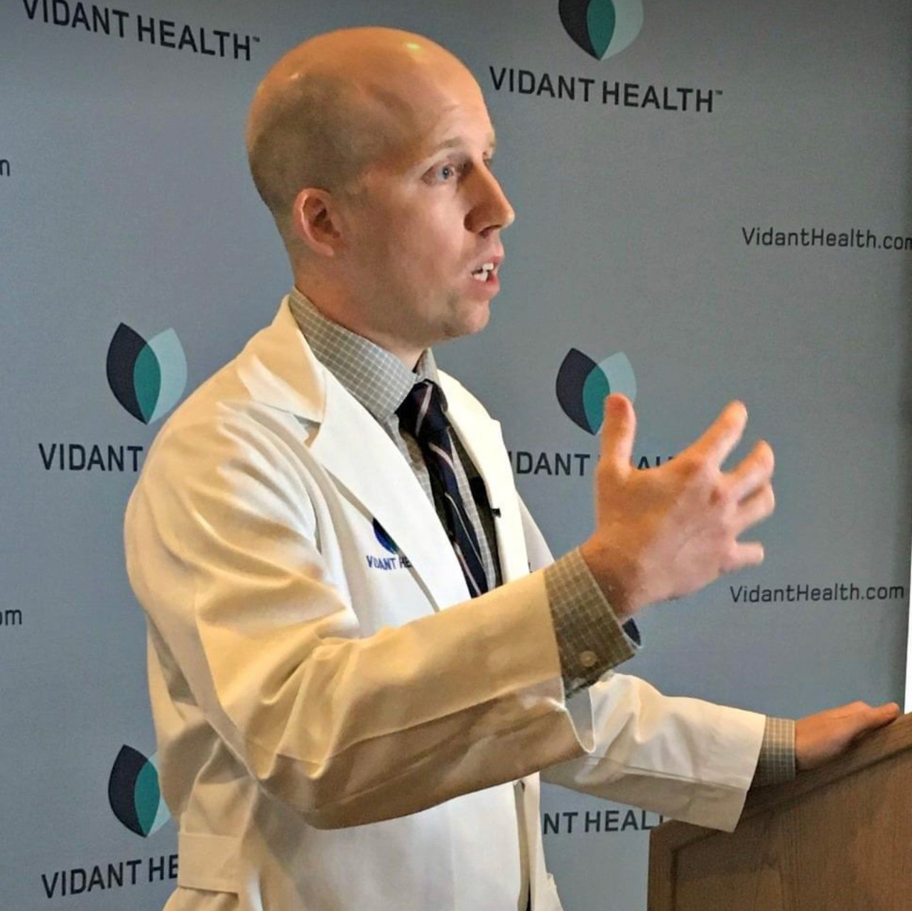
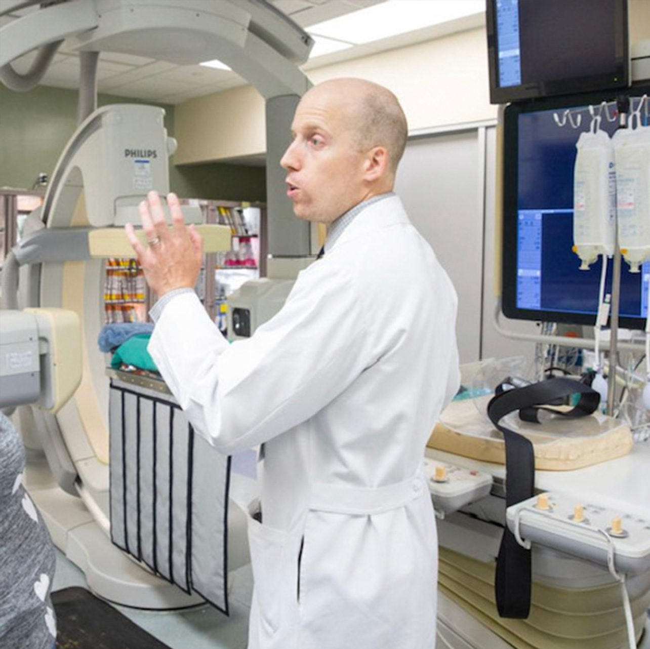
Dr. Dalyai
In The News
Brain Hemorrhage Threatens 8-year-old Boy’s Ability to Walk, Talk or See Again
"Blake was very sick, and there was a high likelihood that this would be a life-threatening hemorrhage,” said Dr. Dalyai. “It was deep in his brain, and it was filling up the fluid pockets, the ventricles that we have in our brain. And that was Blake’s initial extreme injury that needed to be fixed emergently."
Read Full Article